Product Details
| Species Reactivity |
Mouse (Mus musculus) |
| UniProt |
Q5SNZ0 |
| Abbreviation |
CCDC88A |
| Alternative Names |
APE; DKFZp686D0630; GIRDIN; GIV; GRDN; HkRP1; KIAA1212; AKT-phosphorylation enhancer|Galpha-interacting vesicle-associated protein|G{alpha}-interacting vesicle-associated protein|Hook-related protei |
| Range |
Request Information |
| Sensitivity |
Request Information |
| Sample Type |
Serum, Plasma, Other biological fluids |
| Detection Method |
Sandwich |
| Analysis Method |
Quantitive |
| Assay Duration |
1-4.5h |
| Sample Volume |
�1�-�2�0�0� μ�L |
| Detection Wavelengt |
450 nm |
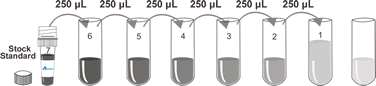
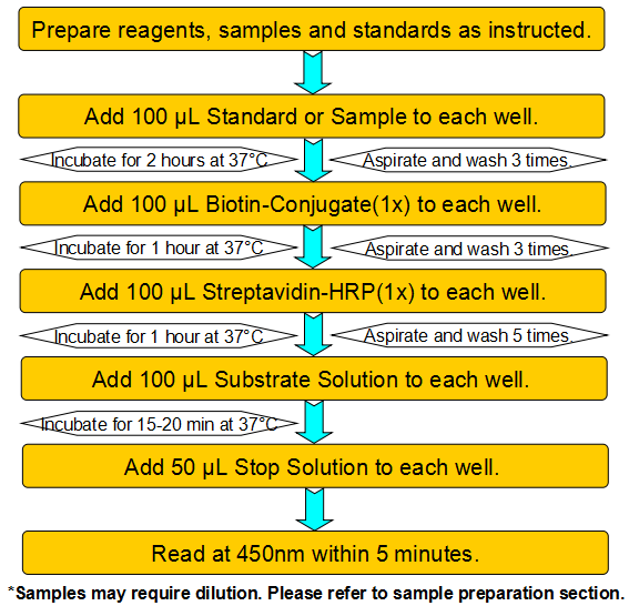
Test principle
This assay employs a two-site sandwich ELISA to quantitate CCDC88A in samples. An antibody specific for CCDC88A has been pre-coated onto a microplate. Standards and samples are pipetted into the wells and any CCDC88A present is bound by the immobilized antibody. After removing any unbound substances, a biotin-conjugated antibody specific for CCDC88A is added to the wells. After washing, Streptavidin conjugated Horseradish Peroxidase (HRP) is added to the wells. Following a wash to remove any unbound avidin-enzyme reagent, a substrate solution is added to the wells and color develops in proportion to the amount of CCDC88A bound in the initial step. The color development is stopped and the intensity of the color is measured.
Product Overview
The deduced 1,870-amino acid protein has a calculated molecular mass of 220 kD. Girdin contains 135 continuous heptad repeats typical of alpha-helical coiled-coil proteins. Northern blot analysis detected variable expression of a 9.5-kb transcript in all tissues examined. Western blot analysis of human brain and testis lysates detected girdin at an apparent molecular mass of 250 kD. Endogenous monkey girdin formed oligomers in COS-7 cells, and mutation analysis indicated that its N-terminal domains facilitated oligomerization.GIV also has several putative peroxisomal targeting signals within the coiled-coil domain and an ATP/GTP-binding site within the C-terminal domain. Western blot analysis detected GIV at an apparent molecular mass of 200 kD in several rat tissues and in all cell lines examined, including human cell lines.
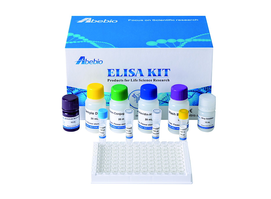
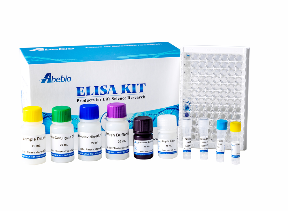
Components
Reagents |
Quantity |
Reagents |
Quantity |
Assay plate (96 Wells) |
1 |
Instruction manual |
1 |
Standard (lyophilized) |
2 |
Sample Diluent |
1 x 20 mL |
Biotin-Conjugate (concentrate 100 x) |
1 x 120 μL |
Biotin-Conjugate Diluent |
1 x 12 mL |
Streptavidin-HRP (concentrate 100 x) |
1 x 120 μL |
Streptavidin-HRP Diluent |
1 x 12 mL |
Wash Buffer (concentrate 25 x) |
1 x 20 mL |
Substrate Solution |
1 x 10 mL |
Stop Solution |
1 x 6 mL |
Adhesive Films |
4 |
Specificity
This assay has high sensitivity and excellent specificity for detection of Mouse CCDC88A. No significant cross-reactivity or interference between Mouse CCDC88A and analogues was observed.
Recovery
Matrices listed below were spiked with certain level of recombinant Mouse CCDC88A and the recovery rates were calculated by comparing the measured value to the expected amount of Mouse CCDC88A in samples.
Precision
Intra-assay Precision (Precision within an assay)
Three samples of known concentration were tested twenty times on one plate to assess intra-assay precision.
Inter-assay Precision (Precision between assays)
Three samples of known concentration were tested in forty separate assays to assess inter-assay precision.
CV (%) = SD/meanX100
Intra-Assay: CV<8%
Inter-Assay: CV<12%
Linearity
The linearity of the kit was assayed by testing samples spiked with appropriate concentration of Mouse CCDC88A and their serial dilutions. The results were demonstrated by the percentage of calculated concentration to the expected.
Stability
The stability of ELISA kit is determined by the loss rate of activity. The loss rate of this kit is less than 5% within the expiration date under appropriate storage condition.
The loss rate was determined by accelerated thermal degradation test. Keep the kit at 37°C for 4 and 7 days, and compare O.D.values of the kit kept at 37°C with that of at recommended temperature. (referring from China Biological Products Standard, which was calculated by the Arrhenius equation. For ELISA kit, 4 days storage at 37°C can be considered as 6 months at 2 - 8°C, which means 7 days at 37°C equaling 12 months at 2 - 8°C).
Sample collection and storage
�S�e�r�u�m�:� �U�s�e� �a� �s�e�r�u�m� �s�e�p�a�r�a�t�o�r� �t�u�b�e� �(�S�S�T�)� �a�n�d� �a�l�l�o�w� �s�a�m�p�l�e�s� �t�o� �c�l�o�t� �f�o�r� �t�w�o� �h�o�u�r�s� �a�t� �r�o�o�m� �t�e�m�p�e�r�a�t�u�r�e� �o�r� �o�v�e�r�n�i�g�h�t� �a�t� �2� �-� �8°�C� �b�e�f�o�r�e� �c�e�n�t�r�i�f�u�g�a�t�i�o�n� �f�o�r� �1�5� �m�i�n�u�t�e�s� �a�t� �1�0�0�0� ×� �g�.� �R�e�m�o�v�e� �s�e�r�u�m� �a�n�d� �a�s�s�a�y� �i�m�m�e�d�i�a�t�e�l�y� �o�r� �a�l�i�q�u�o�t� �a�n�d� �s�t�o�r�e� �s�a�m�p�l�e�s� �a�t� ≤� �-�2�0°�C�.� �A�v�o�i�d� �r�e�p�e�a�t�e�d� �f�r�e�e�z�e�-�t�h�a�w� �c�y�c�l�e�s�.�
�P�l�a�s�m�a�:� �C�o�l�l�e�c�t� �p�l�a�s�m�a� �u�s�i�n�g� �E�D�T�A�,� �o�r� �h�e�p�a�r�i�n� �a�s� �a�n� �a�n�t�i�c�o�a�g�u�l�a�n�t�.� �C�e�n�t�r�i�f�u�g�e� �f�o�r� �1�5� �m�i�n�u�t�e�s� �a�t� �1�0�0�0� ×� �g� �a�t� �2� �-� �8°�C� �w�i�t�h�i�n� �3�0� �m�i�n�u�t�e�s� �o�f� �c�o�l�l�e�c�t�i�o�n�.� �A�s�s�a�y� �i�m�m�e�d�i�a�t�e�l�y� �o�r� �a�l�i�q�u�o�t� �a�n�d� �s�t�o�r�e� �s�a�m�p�l�e�s� �a�t� ≤� �-�2�0°�C�.� �A�v�o�i�d� �r�e�p�e�a�t�e�d� �f�r�e�e�z�e�-�t�h�a�w� �c�y�c�l�e�s�.�
�O�t�h�e�r� �b�i�o�l�o�g�i�c�a�l� �f�l�u�i�d�s�:� �C�e�n�t�r�i�f�u�g�e� �s�a�m�p�l�e�s� �f�o�r� �2�0� �m�i�n�u�t�e�s� �a�t� �1�0�0�0� ×� �g�.� �R�e�m�o�v�e� �p�a�r�t�i�c�u�l�a�t�e�s� �a�n�d� �a�s�s�a�y� �i�m�m�e�d�i�a�t�e�l�y� �o�r� �s�t�o�r�e� �s�a�m�p�l�e�s� �i�n� �a�l�i�q�u�o�t� �a�t� �-�2�0°�C� �o�r� �-�8�0°�C�.� �A�v�o�i�d� �r�e�p�e�a�t�e�d� �f�r�e�e�z�e�/�t�h�a�w� �c�y�c�l�e�s�.
Kits storage instructions
Store at 2-8°C. Please refer to Instruction Manual.

 Phone:
  E-mail:
  Address: